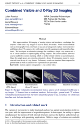
Combined Visible and X-Ray 3D Imaging
Julien Pansiot, Lionel Reveret, and Edmond Boyer
MIUA 2014
Abstract
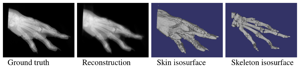
This paper considers 3D imaging of moving objects and introduces a technique that exploits visible and x-ray images to recover dense 3D models. While recent methods such as tomography from cone-beam x-ray can advantageously replace more expensive and higher-dose CT scanners, they still require specific equipment and immobilised patients. We investigate an alternative strategy that combines a single x-ray source and a set of colour cameras to capture rigidly moving samples. The colour cameras allow for coarse marklerless motion tracking, which is further refined with the x-ray information. Once the sample poses are correctly estimated, a dense 3D attenuation model is reconstructed from the set of x-ray frames. Preliminary results on simulated data compared to ground-truth as well as actual in-vivo experiments are presented.
Files
 Paper |
 Presentation |
Bibtex reference
@inproceedings{pansiot14xrays,
    author     =  {Julien Pansiot and Lionel Reveret and Edmond Boyer},
    title      =  {Combined Visible and X-Ray {3D} Imaging},
    booktitle  =  {Medical Image Understanding and Analysis (MIUA)},
    year       =  2014,
    month      =  Jul,
    pages      =  "13--18",
    address    =  {London},
    url        =  "http://www.city.ac.uk/medical-image-understanding-and-analysis-2014/programme",
    eprint     =  "http://julien.pansiot.org/papers/2014_Pansiot_MIUA_Xrays.pdf"
}